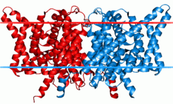
Chloride channel
 Clc chloride channel | |||||||||||
| Identifiers | |||||||||||
|---|---|---|---|---|---|---|---|---|---|---|---|
| Symbol | Voltage_CLC | ||||||||||
| Pfam | PF00654 | ||||||||||
| InterPro | IPR014743 | ||||||||||
| SCOP2 | 1kpl / SCOPe / SUPFAM | ||||||||||
| TCDB | 1.A.11 | ||||||||||
| OPM superfamily | 10 | ||||||||||
| OPM protein | 1ots | ||||||||||
| CDD | cd00400 | ||||||||||
| |||||||||||
Chloride channels are a superfamily of poorly understood ion channels for chloride. These channels may conduct many different ions, but are named for chloride because its concentration in vivo is much higher than other anions.[1] Several families of voltage-gated channels and ligand-gated channels (e.g., the CaCC families) have been characterized in humans.
Voltage-gated chloride channels display a variety of important physiological and cellular roles that include regulation of pH, volume homeostasis, organic solute transport, cell migration, cell proliferation and differentiation. Based on sequence homology the chloride channels can be subdivided into a number of groups.
General functions
Voltage-gated chloride channels are important for setting cell resting membrane potential and maintaining proper cell volume. These channels conduct Cl− as well as other anions such as HCO3−, I−, SCN−, and NO3−. The structure of these channels are not like other known channels. The chloride channel subunits contain between 1 and 12 transmembrane segments. Some chloride channels are activated only by voltage (i.e., voltage-gated), while others are activated by Ca2+, other extracellular ligands, or pH.[2]
CLC family
The CLC family of chloride channels contains 10 or 12 transmembrane helices. Each protein forms a single pore. It has been shown that some members of this family form homodimers. In terms of primary structure, they are unrelated to known cation channels or other types of anion channels. Three CLC subfamilies are found in animals. CLCN1 is involved in setting and restoring the resting membrane potential of skeletal muscle, while other channels play important parts in solute concentration mechanisms in the kidney.[3] These proteins contain two CBS domains. Chloride channels are also important for maintaining safe ion concentrations within plant cells.[4]
Structure and mechanism
The CLC channel structure has not yet been resolved by x-ray crystallography, however the structure of the CLC exchangers has been. Because the primary structure of the channels and exchangers are so similar, most assumptions about the structure of the channels are based on the structure established for the bacterial exchangers.[5]

Each channel or exchanger is composed of two similar subunits—a dimer—each subunit containing one pore. The proteins are formed from two copies of the same protein—a homodimer—though scientists have artificially combined subunits from different channels to form heterodimers. Each subunit binds ions independently of the other, meaning conduction or exchange occur independently in each subunit.[3]

Each subunit consists of two related halves oriented in opposite directions, forming an ‘antiparallel’ structure. These halves come together to form the anion pore.[5] The pore has a filter through which chloride and other anions can pass, but lets little else through. These water-filled pores filter anions via three binding sites—Sint, Scen, and Sext—which bind chloride and other anions. The names of these binding sites correspond to their positions within the membrane. Sint is exposed to intracellular fluid, Scen lies inside the membrane or in the center of the filter, and Sext is exposed to extracellular fluid.[4] Each binding site binds different chloride anions simultaneously. In the exchangers, these chloride ions do not interact strongly with one another, due to compensating interactions with the protein. In the channels, the protein does not shield chloride ions at one binding site from the neighboring negatively charged chlorides.[6] Each negative charge exerts a repulsive force on the negative charges next to it. Researchers have suggested that this mutual repulsion contributes to the high rate of conduction through the pore.[5]
CLC transporters shuttle H+ across the membrane. The H+ pathway in CLC transporters utilizes two glutamate residues—one on the extracellular side, Gluex, and one on the intracellular side, Gluin. Gluex also serves to regulate chloride exchange between the protein and extracellular solution. This means that the chloride and the proton share a common pathway on the extracellular side, but diverge on the intracellular side.[6]
CLC channels also have dependence on H+, but for gating rather than Cl- exchange. Instead of utilizing gradients to exchange two Cl- for one H+, the CLC channels transport one H+ while simultaneously transporting millions of anions.[6] This corresponds with one cycle of the slow gate.
Eukaryotic CLC channels also contain cytoplasmic domains. These domains have a pair of CBS motifs, whose function is not fully characterized yet.[5] Though the precise function of these domains is not fully characterized, their importance is illustrated by the pathologies resulting from their mutation. Thomsen’s disease, Dent’s disease, infantile malignant osteopetrosis, and Bartter’s syndrome are all genetic disorders due to such mutations.
At least one role of the cytoplasmic CBS domains regards regulation via adenosine nucleotides. Particular CLC transporters and proteins have modulated activity when bound with ATP, ADP, AMP, or adenosine at the CBS domains. The specific effect is unique to each protein, but the implication is that certain CLC transporters and proteins are sensitive to the metabolic state of the cell.[6]
Selectivity
The Scen acts as the primary selectivity filter for most CLC proteins, allowing the following anions to pass through, from most selected to least selected: SCN-, Cl-, Br-, NO3-, I-. Altering a serine residue at the selectivity filter, labeled Sercen, to a different amino acid alters the selectivity.[6]
Gating and kinetics
Gating occurs through two mechanisms: protopore or fast gating and common or slow gating. Common gating involves both protein subunits closing their pores at the same time (cooperation), while protopore gating involves independent opening and closing of each pore.[5] As the names imply, fast gating occur at a much faster rate than slow gating. Precise molecular mechanisms for gating are still being studied.
For the channels, when the slow gate is closed, no ions permeate through the pore. When the slow gate is open, the fast gates open spontaneously and independently of one another. Thus, the protein could have both gates open, or both gates closed, or just one of the two gates open. Single-channel patch-clamp studies demonstrated this biophysical property even before the dual-pore structure of CLC channels had been resolved. Each fast gate opens independently of the other and the ion conductance measured during these studies reflects a binomial distribution.[3]
H+ transport promotes opening of the common gate in CLC channels. For every opening and closing of the common gate, one H+ is transported across the membrane. The common gate is also affected by the bonding of adenosine nucleotides to the intracellular CBS domains. Inhibition or activation of the protein by these domains is specific to each protein.[6]
Function
The CLC channels allow chloride to flow down its electrochemical gradient, when open. These channels are expressed on the cell membrane. CLC channels contribute to the excitability of these membranes as well as transport ions across the membrane.[3]
The CLC exchangers are localized to intracellular components like endosomes or lysosomes and help regulate the pH of their compartments.[3]
Pathology
Bartter's syndrome, which is associated with renal salt wasting and hypokalemic alkalosis, is due to the defective transport of chloride ions and associated ions in the thick ascending loop of Henle. CLCNKB has been implicated.[7]
Another inherited disease that affects the kidney organs is Dent's Disease, characterised by low molecular weight proteinuria and hypercalciuria where mutations in CLCN5 are implicated.[7]
Thomsen disease is associated with dominant mutations and Becker disease with recessive mutations in CLCN1.[7]
Genes
CFTR
CFTR is a chloride channel belonging to the superfamily of ABC transporters. Each channel has two transmembrane domains and two nucleotide binding domains. ATP binding to both nucleotide binding domains causes changes these domains to associate, further causing changes that open up the ion pore. When ATP is hydrolyzed, the nucleotide binding domains dissociate again and the pore closes.[8]
Pathology
Cystic fibrosis is caused by mutations in the CFTR gene, which prevents the proper folding of the protein and subsequent degradation, resulting in decreased numbers of chloride channels in the body. This causes the buildup of mucus in the body and chronic infections.[8]
Other chloride channels and families
- CLIC family
- GABAA
- Glycine Receptor
- Calcium-activated chloride channel
Commercial applications
This section may be confusing or unclear to readers. (February 2016) |
Some organic materials disrupt chloride channels in fleas, causing death. Selamectin is the active ingredient in Revolution, a topical insecticide and antihelminthic used on dogs and cats. Selamectin works by replacing glutamate which normally interacts with receptors that open chloride channels at muscle synapses found in parasites. Unlike glutamate, selamectin activates the chloride current without desensitization, thereby producing prolonged hyperpolarization and impaired muscle contraction.
References
- ^ Jentsch, Thomas J.; Stein, Valentin; Weinreich, Frank; Zdebik, Anselm A. (2002-04-01). "Molecular Structure and Physiological Function of Chloride Channels". Physiological Reviews. 82 (2): 503–568. doi:10.1152/physrev.00029.2001. ISSN 0031-9333. PMID 11917096.
- ^ "Diversity of Cl− Channels". Cell. Mol. Life Sci. 63 (1): 12–24. January 2006. doi:10.1007/s00018-005-5336-4. PMC 2792346. PMID 16314923.
{{cite journal}}: Unknown parameter|authors=ignored (help) - ^ a b c d e Stölting, Gabriel; Fischer, Martin; Fahlke, Christoph (2014-01-01). "CLC channel function and dysfunction in health and disease". Membrane Physiology and Membrane Biophysics. 5: 378. doi:10.3389/fphys.2014.00378. PMC 4188032. PMID 25339907.
{{cite journal}}: CS1 maint: unflagged free DOI (link) - ^ "Tonoplast-located GmCLC1 and GmNHX1 from soybean enhance NaCl tolerance in transgenic bright yellow (BY)-2 cells". Plant Cell Environ. 29 (6): 1122–37. June 2006. doi:10.1111/j.1365-3040.2005.01487.x. PMID 17080938.
{{cite journal}}: Unknown parameter|authors=ignored (help) - ^ a b c d e Dutzler, Raimund. "A structural perspective on ClC channel and transporter function". FEBS Letters. 581 (15): 2839–2844. doi:10.1016/j.febslet.2007.04.016.
- ^ a b c d e f Accardi, Alessio; Picollo, Alessandra (2010-08-01). "CLC channels and transporters: Proteins with borderline personalities". Biochimica et Biophysica Acta (BBA) - Biomembranes. 1798 (8): 1457–1464. doi:10.1016/j.bbamem.2010.02.022. PMC 2885512. PMID 20188062.
- ^ a b c Planells-Cases, Rosa; Jentsch, Thomas J. (2009-03-01). "Chloride channelopathies". Biochimica et Biophysica Acta (BBA) - Molecular Basis of Disease. 1792 (3): 173–189. doi:10.1016/j.bbadis.2009.02.002.
- ^ a b Gadsby, David C.; Vergani, Paola; Csanády, László. "The ABC protein turned chloride channel whose failure causes cystic fibrosis". Nature. 440 (7083): 477–483. doi:10.1038/nature04712. PMC 2720541. PMID 16554808.
Further reading
- Schmidt-Rose T, Jentsch TJ (August 1997). "Reconstitution of functional voltage-gated chloride channels from complementary fragments of CLC-1". J. Biol. Chem. 272 (33): 20515–21. doi:10.1074/jbc.272.33.20515. PMID 9252364.
{{cite journal}}: CS1 maint: unflagged free DOI (link) - "Mutations in the human skeletal muscle chloride channel gene (CLCN1) associated with dominant and recessive myotonia congenita". Neurology. 47 (4): 993–8. October 1996. doi:10.1212/wnl.47.4.993. PMID 8857733.
{{cite journal}}: Unknown parameter|authors=ignored (help) - "ClC chloride channels". Genome Biol. 2 (2): REVIEWS3003. 2001. doi:10.1186/gb-2001-2-2-reviews3003. PMC 138906. PMID 11182894.
{{cite journal}}: Unknown parameter|authors=ignored (help)CS1 maint: unflagged free DOI (link) - Singh H (2010). "Two decades with dimorphic Chloride Intracellular Channels (CLICs)". FEBS Letters. 584 (10): 2112–21. doi:10.1016/j.febslet.2010.03.013. PMID 20226783.
External links
- Chloride+channels at the U.S. National Library of Medicine Medical Subject Headings (MeSH)
- UMich Orientation of Proteins in Membranes families/superfamily-10 - CLC chloride channels
