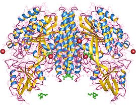
Transferrin receptor
| Transferrin receptor 1 | |||||||
|---|---|---|---|---|---|---|---|
 Transferrin receptor 1, dimer, Human | |||||||
| Identifiers | |||||||
| Symbol | TFRC | ||||||
| Alt. symbols | CD71, TFR1 | ||||||
| NCBI gene | 7037 | ||||||
| HGNC | 11763 | ||||||
| OMIM | 190010 | ||||||
| RefSeq | NM_003234 | ||||||
| UniProt | P02786 | ||||||
| Other data | |||||||
| Locus | Chr. 3 q29 | ||||||
| |||||||
| Transferrin receptor 2 | |||||||
|---|---|---|---|---|---|---|---|
| Identifiers | |||||||
| Symbol | TFR2 | ||||||
| Alt. symbols | HFE3, TFRC2 | ||||||
| NCBI gene | 7036 | ||||||
| HGNC | 11762 | ||||||
| OMIM | 604720 | ||||||
| RefSeq | NM_003227 | ||||||
| UniProt | Q9UP52 | ||||||
| Other data | |||||||
| Locus | Chr. 7 q22 | ||||||
| |||||||
Transferrin receptor (TfR) is a carrier protein for transferrin. It is needed for the import of iron into the cell and is regulated in response to intracellular iron concentration. It imports iron by internalizing the transferrin-iron complex through receptor-mediated endocytosis.[1] The existence of a receptor for transferrin iron uptake had been recognized over half a century back.[2] Earlier two transferrin receptors in humans, transferrin receptor 1 and transferrin receptor 2 had been characterized and until recently cellular iron uptake was believed to occur chiefly via these two well documented transferrin receptors. Both these receptors are transmembrane, glycoproteins. TfR1 is a high affinity ubiquitously expressed receptor while expression of TfR2 is restricted to certain cell types and is unaffected by intracellular iron concentrations. TfR2 binds to transferrin with a 25-30 fold lower affinity than TfR1.[3][4] Although TfR1 mediated iron uptake is the major pathway for iron acquisition by most cells and especially developing erythrocytes, several studies have indicated that the uptake mechanism varies depending upon the cell type. It is also reported that Tf uptake exists independent of these TfRs although the mechanisms are not well characterized.[5][6][7][8] The multifunctional glycolytic enzyme glyceraldehyde 3-phosphate dehydrogenase (GAPDH,EC 1.2.1.12) has been shown to utilize post translational modifications to exhibit higher order moonlighting behavior wherein it switches its function as a holo or apo transferrin receptor leading to either iron delivery or iron export respectively.[9][10][11]
Translational regulation
Low iron concentrations promote increased levels of transferrin receptor, to increase iron intake into the cell. Thus, transferrin receptor maintains cellular iron homeostasis.
TfR production in the cell is regulated according to iron levels by iron-responsive element-binding proteins, IRP1 and IRP2. In the absence of iron, one of these proteins (generally IRP2) binds to the hairpin like structure (IRE) that is in the 3' UTR of the TfR mRNA. Once binding occurs, the mRNA is stabilized and degradation is inhibited.
See also
References
- ^ Qian ZM, Li H, Sun H, Ho K (December 2002). "Targeted drug delivery via the transferrin receptor-mediated endocytosis pathway". Pharmacological Reviews. 54 (4): 561–87. doi:10.1124/pr.54.4.561. PMID 12429868.; Figure 3: The cycle of transferrin and transferrin receptor 1-mediated cellular iron uptake.
- ^ Jandl JH, Inman JK, Simmons RL, Allen DW (January 1959). "Transfer of iron from serum iron-binding protein to human reticulocytes". The Journal of Clinical Investigation. 38 (1, Part 1): 161–85. doi:10.1172/JCI103786. PMC 444123. PMID 13620780.
- ^ Kawabata H, Germain RS, Vuong PT, Nakamaki T, Said JW, Koeffler HP (June 2000). "Transferrin receptor 2-alpha supports cell growth both in iron-chelated cultured cells and in vivo". The Journal of Biological Chemistry. 275 (22): 16618–25. doi:10.1074/jbc.M908846199. PMID 10748106.
{{cite journal}}: CS1 maint: unflagged free DOI (link) - ^ West AP, Bennett MJ, Sellers VM, Andrews NC, Enns CA, Bjorkman PJ (December 2000). "Comparison of the interactions of transferrin receptor and transferrin receptor 2 with transferrin and the hereditary hemochromatosis protein HFE". The Journal of Biological Chemistry. 275 (49): 38135–8. doi:10.1074/jbc.C000664200. PMID 11027676.
{{cite journal}}: CS1 maint: unflagged free DOI (link) - ^ Gkouvatsos K, Papanikolaou G, Pantopoulos K (March 2012). "Regulation of iron transport and the role of transferrin". Biochimica et Biophysica Acta. 1820 (3): 188–202. doi:10.1016/j.bbagen.2011.10.013. PMID 22085723.
- ^ Trinder D, Zak O, Aisen P (June 1996). "Transferrin receptor-independent uptake of differic transferrin by human hepatoma cells with antisense inhibition of receptor expression". Hepatology. 23 (6): 1512–20. doi:10.1053/jhep.1996.v23.pm0008675172. PMID 8675172.
- ^ Kozyraki R, Fyfe J, Verroust PJ, Jacobsen C, Dautry-Varsat A, Gburek J, Willnow TE, Christensen EI, Moestrup SK (October 2001). "Megalin-dependent cubilin-mediated endocytosis is a major pathway for the apical uptake of transferrin in polarized epithelia". Proceedings of the National Academy of Sciences of the United States of America. 98 (22): 12491–6. doi:10.1073/pnas.211291398. PMC 60081. PMID 11606717.
- ^ Yang J, Goetz D, Li JY, Wang W, Mori K, Setlik D, Du T, Erdjument-Bromage H, Tempst P, Strong R, Barasch J (November 2002). "An iron delivery pathway mediated by a lipocalin". Molecular Cell. 10 (5): 1045–56. doi:10.1016/s1097-2765(02)00710-4. PMID 12453413.
- ^ Sirover MA (December 2014). "Structural analysis of glyceraldehyde-3-phosphate dehydrogenase functional diversity". The International Journal of Biochemistry & Cell Biology. 57: 20–6. doi:10.1016/j.biocel.2014.09.026. PMC 4268148. PMID 25286305.
- ^ Boradia VM, Raje M, Raje CI (December 2014). "Protein moonlighting in iron metabolism: glyceraldehyde-3-phosphate dehydrogenase (GAPDH)". Biochemical Society Transactions. 42 (6): 1796–801. doi:10.1042/BST20140220. PMID 25399609.
- ^ Sheokand N, Malhotra H, Kumar S, Tillu VA, Chauhan AS, Raje CI, Raje M (October 2014). "Moonlighting cell-surface GAPDH recruits apotransferrin to effect iron egress from mammalian cells". Journal of Cell Science. 127 (Pt 19): 4279–91. doi:10.1242/jcs.154005. PMID 25074810.
Further reading
- Testa U, Kühn L, Petrini M, Quaranta MT, Pelosi E, Peschle C (July 1991). "Differential regulation of iron regulatory element-binding protein(s) in cell extracts of activated lymphocytes versus monocytes-macrophages". The Journal of Biological Chemistry. 266 (21): 13925–30. PMID 1856222.
- Daniels TR, Delgado T, Rodriguez JA, Helguera G, Penichet ML (November 2006). "The transferrin receptor part I: Biology and targeting with cytotoxic antibodies for the treatment of cancer". Clinical Immunology. 121 (2): 144–58. doi:10.1016/j.clim.2006.06.010. PMID 16904380.; Figure 3: Cellular uptake of iron through the Tf system via receptor-mediated endocytosis.
- Daniels TR, Delgado T, Helguera G, Penichet ML (November 2006). "The transferrin receptor part II: targeted delivery of therapeutic agents into cancer cells". Clinical Immunology. 121 (2): 159–76. doi:10.1016/j.clim.2006.06.006. PMID 16920030.
External links
- Transferrin+receptor at the U.S. National Library of Medicine Medical Subject Headings (MeSH)
- Okam M (2001-01-29). "Transerrin and Iron Transport Physiology". Information Center for Sickle Cell and Thalassemic Disorders. Brigham and Women's Hospital and Harvard Medical School. Retrieved 2010-12-19.
